The winners of Nikon Instruments Inc.’s 50th anniversary Nikon Small World Photomicrography Competition were revealed. About 2,100 amateurs, researchers, students, and other scientists from 80 nations entered this year’s famous photomicrography competition.
Established in 1974, the Nikon Small World competition is regarded as the premier venue for honouring the talent, expertise, technical mastery, and photographic brilliance required for photomicrography—the process of taking pictures under a microscope. The Nikon Small World in Motion contest for video micrography, which announced its winners last month, has joined the Nikon Small World 2024 competition.
This year’s winner is Dr. Bruno Cisterna, who worked with Dr. Eric Vitriol of Augusta University to create the amazing image of differentiated mouse brain tumour cells that displays the nuclei, microtubules, and actin cytoskeleton. This illustration demonstrates how devastating neurological conditions like Alzheimer’s and ALS can result from abnormalities in the cytoskeleton, the cell’s basic foundation.
In addition to being aesthetically pleasing, Dr. Cisterna’s research showed that the protein profilin 1 (PFN1), which is essential for constructing the cell’s structure, also contributes to the upkeep of microtubules, which are like tiny cellular highways and crucial for cellular transport.
“When PFN1 or related processes are disrupted, these highways can malfunction, leading to cellular damage similar to what is observed in neurodegenerative diseases,” Nikon Instruments explains.
“One of the main problems with neurodegenerative diseases is that we don’t fully understand what causes them,” says Dr. Cisterna. “To develop effective treatments, we need to figure out the basics first. Our research is crucial for uncovering this knowledge and ultimately finding a cure. Differentiated cells could be used to study how mutations or toxic proteins that cause Alzheimer’s or ALS alter neuronal morphology, as well as to screen potential drugs or gene therapies aimed at protecting neurons or restoring their function.”
Since Dr. Cisterna spent almost three months “perfecting the staining process to ensure clear visibility of the cells,” his patience and ability were greatly needed to take this picture.
“After allowing five days for the cells to differentiate, I had to find the right field of view where the differentiated and non-differentiated cells interacted. This took about three hours of precise observation under the microscope to capture the right moment, involving many attempts and countless hours of work to get it just right,” Dr. Cisterna adds.
Along with the wonderful honour of winning the 50th Nikon Small World Photomicrography Contest, Dr. Cisterna and his team’s research, which was published four months ago in the Journal of Cell Biology, may help millions of people suffering from neurodegenerative diseases and lead to a breakthrough in neurological research. Such research has the power to change lives in amazing ways.
Many of the photos entered in this year’s competition, as well as the previous 49 years, show this attitude of scientific advancement.
“At 50 years, Nikon Small World is more than just an imaging competition — it’s become a gallery that pays tribute to the extraordinary individuals who make it possible. They are the driving force behind this event, masterfully blending science and art to reveal the wonders of the microscopic world and what we can learn from it to the public,” says Eric Flem, Senior Manager of CRM and Communications at Nikon Instruments.
“Sometimes, we overlook the tiny details of the world around us. Nikon Small World serves as a reminder to pause, appreciate the power and beauty of the little things, and to cultivate a deeper curiosity to explore and question,” Flem adds.
Second and Third Place Images
For his captivating image of electricity arcing between a wire and a pin, Dr. Marcel Clemens won second prize.
Electrical arc between a pin and a wire | Dr. Marcel Clemens
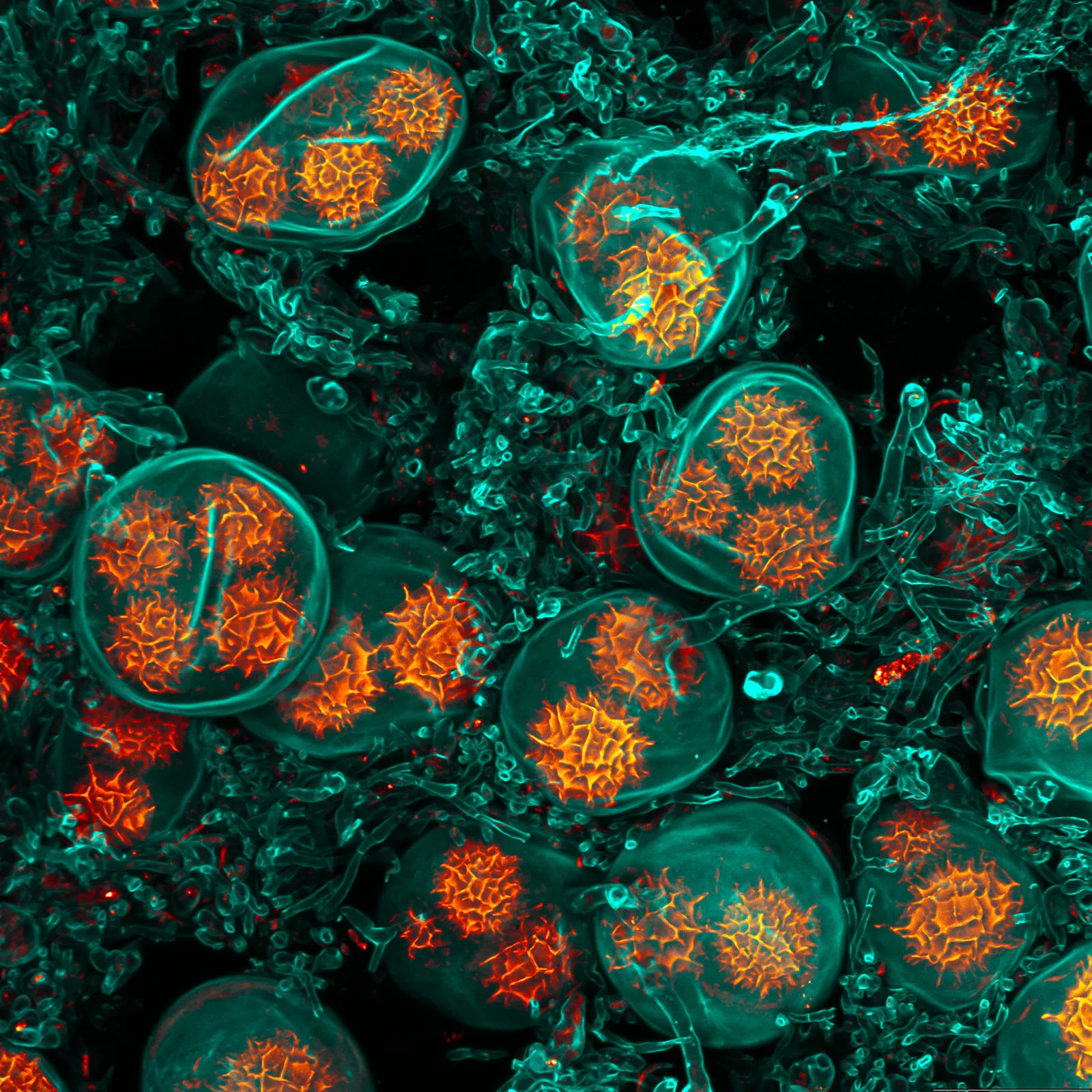
Leaf of a cannabis plant. The bulbous glands are trichomes. The bubbles inside are cannabinoid vesicles | Chris Romaine
The Other Twenty
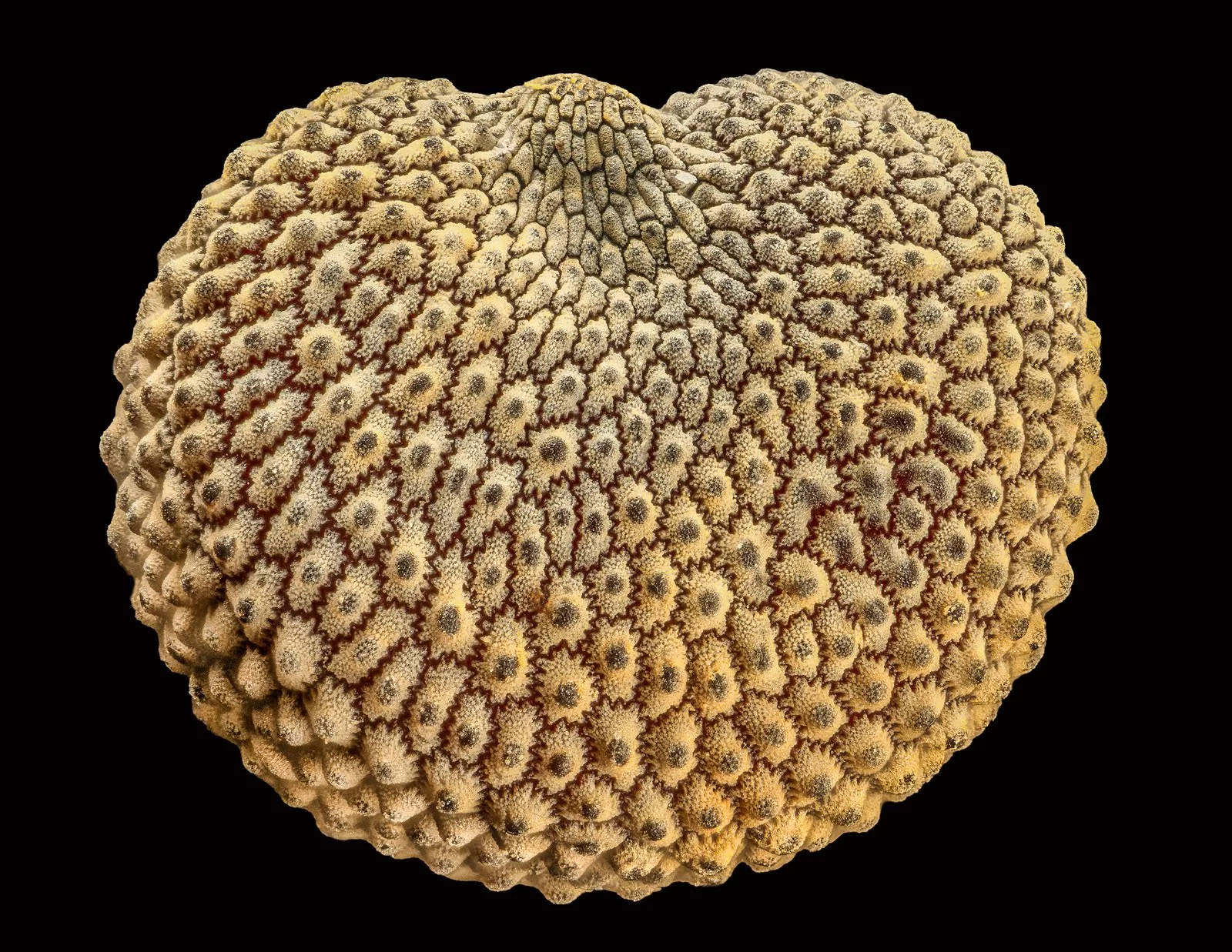
TheNikon Small World Photomicrography Contestwebsite features all 87 of the photographs that were recognised, in addition to the top three winners. The 17 additional photos that make up the top 20 in the Nikon Small World 2024 competition are listed here, arranged from fourth to twentieth.
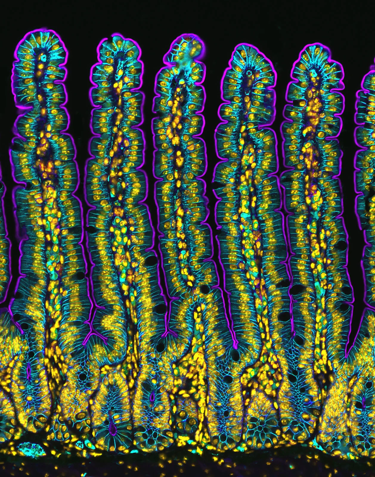
Section of a small intestine of a mouse | Dr. Amy Engevik
Cluster of octopus (Octopus hummelincki) eggs | Thomas Barlow & Connor Gibbons
Slime mold (Cribraria cancellata) | Henri Koskinen
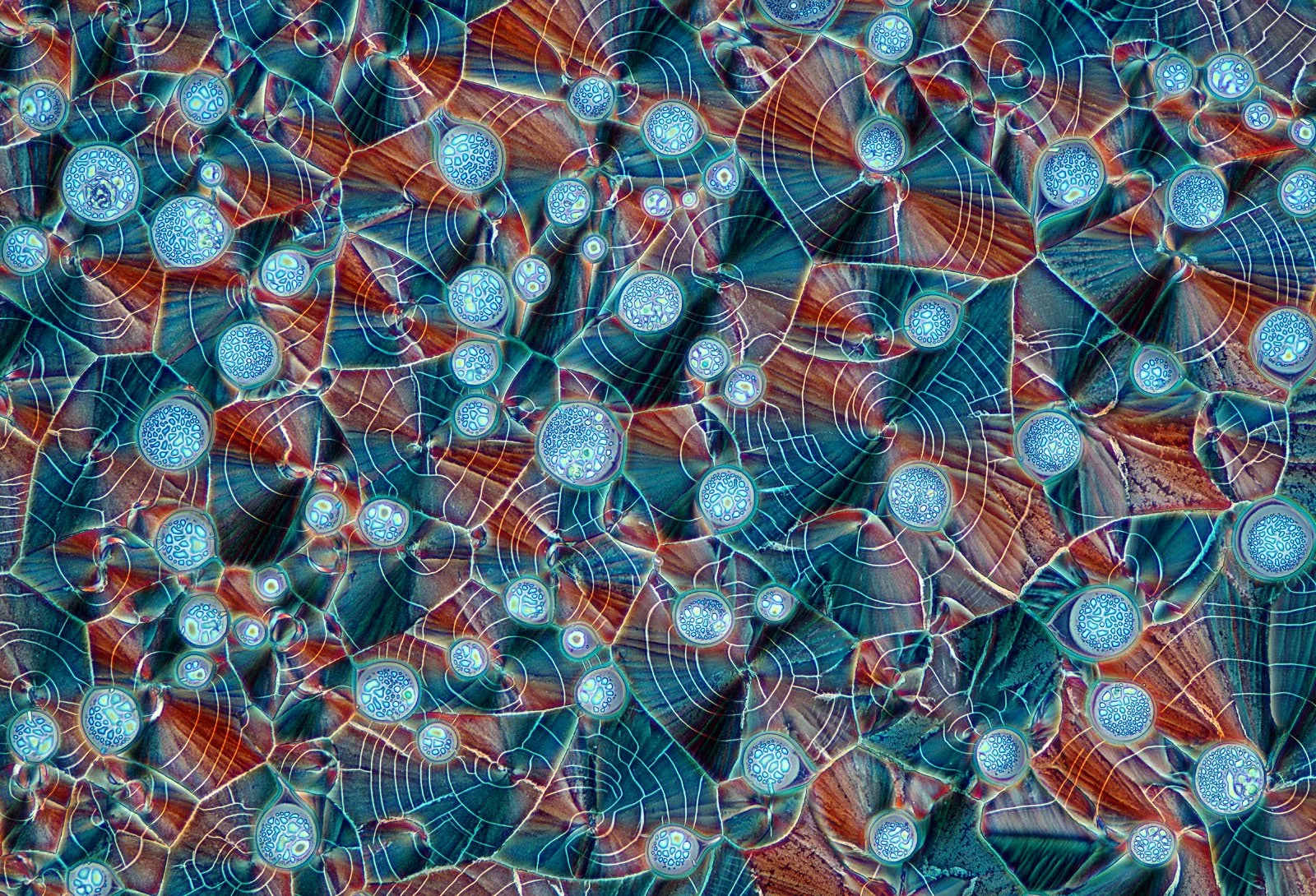
Cross section of European beach grass (Ammophila arenaria) leaf | Gerhard Vlcek
A neuron densely covered in dendritic spines from the striatum of an adult rat brain | Stephanie Huang
Pollen in a garden spider (Araneus) web | John-Oliver Dum
Spores of black truffle (Tuber melanosporum) | Jan Martinek
Slime mold on a rotten twig with water droplets | Dr. Ferenc Halmos
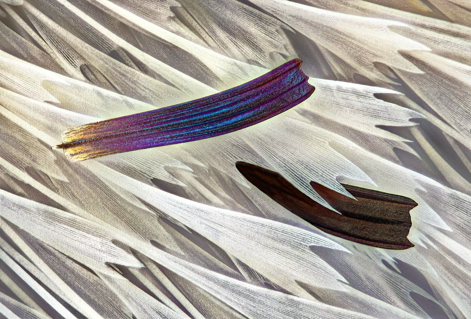
Wing scales of a butterfly (Papilio ulysses) on a medical syringe needle | Daniel Knop
Eyes of green crab spider (Diaea dorsata) | Paweł Błachowicz
Recrystallized mixture of hydroquinone and myoinositol | Marek Miś
Isolated scales on Madagascan sunset moth wing (Chrysiridia ripheus) | Sébastien Malo
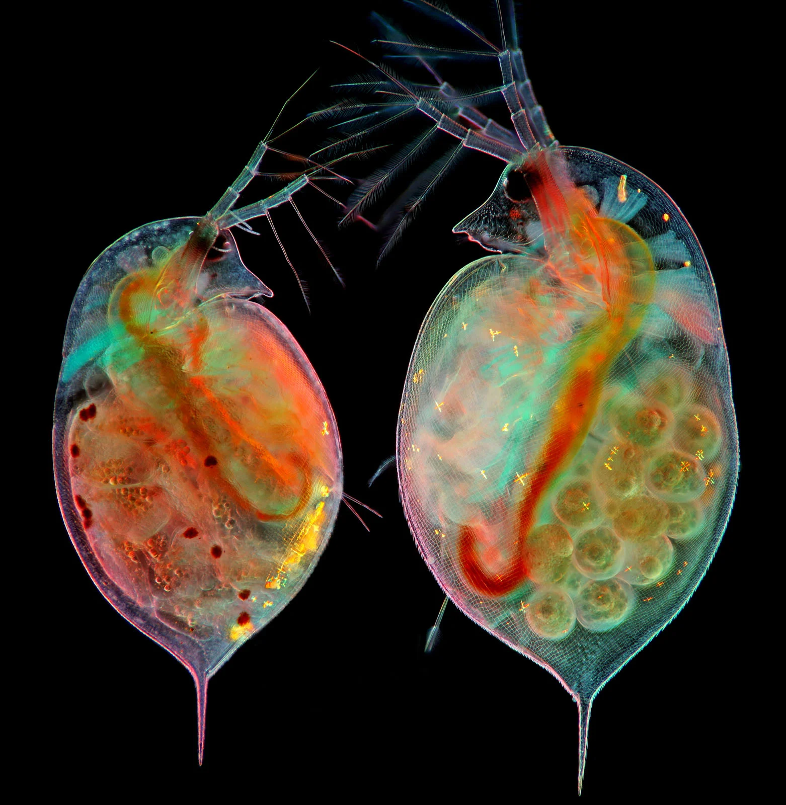
Two water fleas (Daphnia sp.) with embryos (left) and eggs (right) | Marek Miś
Stonewort algae (Chara virgata) reproductive organs — oogonia (female organs) and antheridia (male organs) | Dr. Frantisek Bednar
An insect egg parasitized by a wasp | Alison Pollack
Seed of a Silene plant | Alison Pollack
Early stage of mouse neuroblastoma cell differentiation (actin, microtubules, and mitochondria) | Dr. Bruno Cisterna & Dr. Eric Vitriol
The Nikon Small World website features all of the acknowledged competitors. The competition is amazing, and there are a lot of stunning, intriguing microscope photos to view.
FilmPix Media articles may include affiliate links; if you buy something through such a link, we may earn a commission at no additional cost.
Matteo Bianchi is an Italian media journalist with extensive experience covering photography and videography. He specializes in in-depth gear reviews, industry trends, and technological developments shaping the visual media landscape.